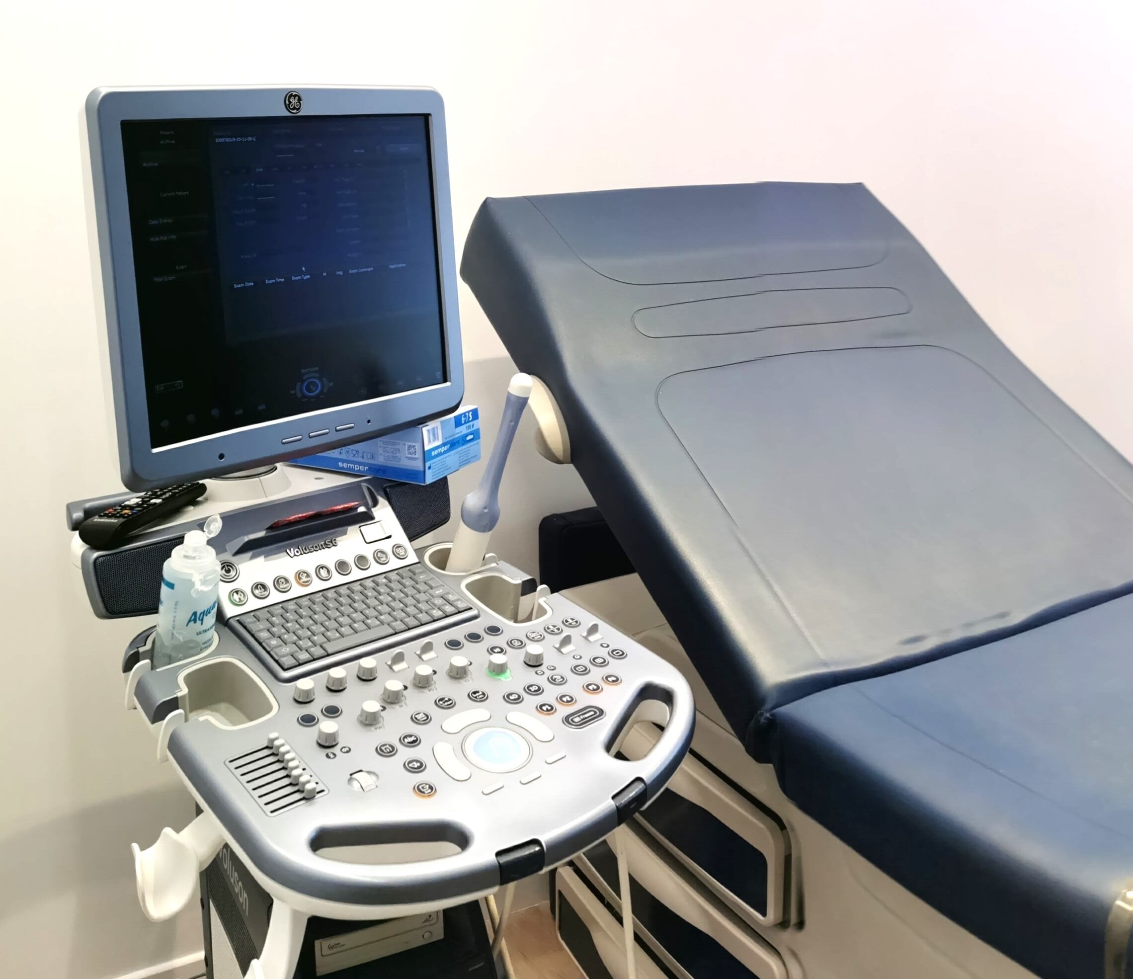
Ultrasound is perhaps the most important diagnostic tool in the daily practice of gynecology and obstetrics. It is a non-invasive, patient-friendly imaging method that helps us see inside our body. Once a year, as part of the annual gynecological check-up, we do an ultrasound of the inner genitals. If we need to monitor the course of a finding, ultrasounds are done more often. We use special probes different each time, depending on the position of the point we are focusing on. In gynecology, the use of the intravaginal probe helps us to check the anatomy of the uterus, to identify congenital abnormalities, polyps, fibroids, … We recognize the ovaries, we check their functionality. We identify possible ovarian cysts and separate them into benign or not, depending on their ultrasound image. We see signs of inflammation, endometriosis, dilated tubes. The use of the 3D intravaginal head gives us images comparable to magnetic resonance imaging in the anatomical variants of the uterus: arcuate, tilted, twin, unicorn.
Within a few minutes we have gathered, without causing discomfort to our patient, completely painlessly, the information we need to help her therapeutically in the problem that concerns her.

